fluoroscopy guided injection
Fluoroscopy guided injection employs real‑time X‑ray imaging to accurately deliver medication into joints or soft tissues. The technique enhances precision, reduces complications, and offers immediate feedback on needle placement and contrast spread. Low‑dose protocols ensure safety and clarity.
Definition and Clinical Context
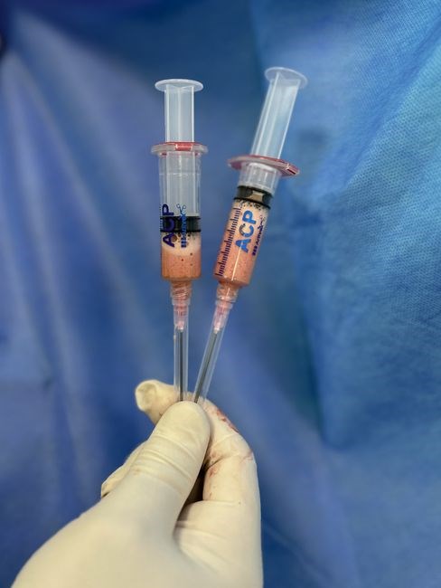
Fluoroscopy guided injection is a minimally invasive procedure that utilizes real‑time X‑ray imaging to direct a needle into a target structure—typically a joint, tendon sheath, or muscle belly—while the operator observes the needle tip, contrast spread, and surrounding anatomy. The technique is widely employed in musculoskeletal medicine to deliver local anesthetics, corticosteroids, hyaluronic acid, or other therapeutic agents with high precision, thereby improving diagnostic accuracy and therapeutic outcomes. In contrast to blind or ultrasound‑guided injections, fluoroscopy offers superior visualization of deep or anatomically complex sites such as the hip, sacroiliac joint, or piriformis muscle, and it allows the clinician to confirm correct placement before medication administration. The procedure is performed in a radiology suite equipped with a C‑arm fluoroscope, lead shielding, and radiation monitoring devices. Patients are positioned to provide optimal access while minimizing radiation exposure, and a low‑dose protocol is employed to keep cumulative dose below established safety thresholds. Fluoroscopy guided injections are indicated for a range of conditions, including osteoarthritis, tendonitis, bursitis, and neuropathic pain syndromes, and they serve both diagnostic and therapeutic purposes. The real‑time feedback provided by fluoroscopy enables immediate adjustment of needle trajectory, reduces the risk of inadvertent intra‑vascular or intra‑organ puncture, and enhances patient confidence by demonstrating procedural accuracy. Overall, fluoroscopy guided injection represents a cornerstone of modern interventional pain management, combining imaging precision with pharmacologic efficacy to deliver targeted relief while maintaining patient safety.

Indications for Fluoroscopy Guided Injection
Fluoroscopy guided injections are indicated for deep joint pain, osteoarthritis, sacroiliac dysfunction, bursitis, and neuropathic conditions where precise needle placement is critical. They provide accurate delivery of medication, reduce complications, and improve outcomes. Patient safety and diagnostic clarity!
Common Musculoskeletal Conditions

Fluoroscopy guided injections are frequently employed for a spectrum of deep‑tissue musculoskeletal disorders that are difficult to access with conventional palpation. The most common indications include osteoarthritic degeneration of the hip, knee, shoulder, and ankle joints, where intra‑articular corticosteroids or hyaluronic acid can be precisely delivered to reduce inflammation and pain. Sacroiliac joint dysfunction, often presenting as low‑back pain radiating to the buttocks, benefits from targeted anesthetic or steroid injections under fluoroscopic visualization to confirm accurate placement. Bursitis of the glenohumeral, subacromial, trochanteric, and iliopsoas bursae is another frequent target, as the needle can be advanced to the bony end point while avoiding neurovascular structures. Neuropathic pain from lumbar radiculopathy or sciatic nerve irritation is addressed through epidural or paravertebral injections, with fluoroscopy ensuring the contrast outlines the epidural space and the drug is delivered adjacent to the affected nerve root. Tendinopathies of the rotator cuff, Achilles, and patellar tendons are also treated, especially when conservative measures fail; the fluoroscope allows the clinician to place the needle within the tendon sheath or the site of tendon degeneration. Finally, injections for rheumatoid arthritis are performed to deliver disease agents into the inflamed joint improving symptom control and preserving joint integrity!!
Equipment and Setup Requirements
Fluoroscope must have high‑resolution flat‑panel detector, adjustable dose‑rate, and real‑time image processing. Sterile field, lead aprons, thyroid shield, and dosimeter are mandatory. Use low‑kV, pulsed mode to minimize exposure while maintaining image quality. Ensure local compliance. now!?
Fluoroscope Specifications and Radiation Safety
Modern fluoroscopes for musculoskeletal injections integrate flat‑panel detectors with 2‑mm spatial resolution, enabling clear delineation of bone and soft‑tissue interfaces. A pulsed‑mode source operating at 60–80 kV and 1–5 mA, combined with automatic exposure control, limits dose to <0.5 mGy per frame. The system’s real‑time image‑processing algorithms sharpen edges while suppressing noise, allowing clinicians to identify the target needle tip with sub‑millimetre accuracy.
Radiation safety hinges on three pillars: dose optimization, shielding, and monitoring. Dose optimization is achieved by selecting the lowest acceptable kV, using pulsed rather than continuous fluoroscopy, and applying image‑reduction filters. Lead aprons (≥0.5 mm Pb equivalent) and thyroid collars protect the operator’s most radiosensitive tissues. A personal dosimeter records cumulative exposure, and the fluoroscope’s built‑in dose‑area product (DAP) meter provides real‑time feedback to the operator.
Compliance with national guidelines (e.g., AAPM TG‑111, ICRP 103) requires annual equipment calibration, regular image‑quality audits, and documentation of all procedures. Training programs should emphasize the ALARA principle, proper positioning of the patient and staff, and the use of pulsed fluoroscopy at the lowest frame rate that still yields diagnostic image quality. By integrating these specifications and safety practices, clinicians can deliver accurate, effective injections while maintaining radiation exposure within acceptable limits.
- Use pulsed fluoroscopy at 5–10 fps.
- Apply image‑reduction filter (0.5–1.0).
- Maintain patient distance of 50–70 cm.
- Verify needle tip with contrast injection.
- Record DAP after each procedure.
Standard Procedure Techniques
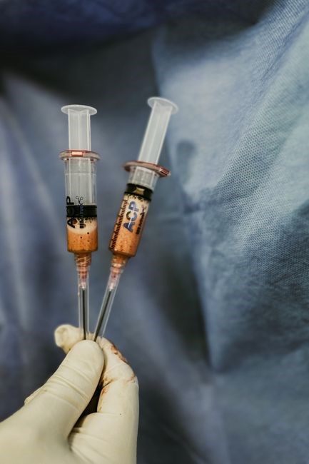
Preparation begins with aseptic skin prep and local anesthesia. The fluoroscope is positioned to visualize the target joint. A sterile needle is advanced under real‑time imaging, and contrast confirms placement before medication injection. Sterile technique is essential. Patient safety is paramount.
Piriformis Muscle Injection
Piriformis muscle injections are performed to relieve sciatic nerve compression and chronic gluteal pain. The procedure begins with patient positioning in lateral decubitus or prone, ensuring the hip is slightly flexed to expose the gluteal region. After sterile preparation, a local anesthetic (1 mL of 1% lidocaine buffered with 1 mL of 8.4 % sodium bicarbonate) is infiltrated superficially to numb the skin and subcutaneous tissues. A 22‑gauge, 5‑inch needle is then introduced at a 45° angle from the lateral aspect of the greater trochanter, targeting the posterior gluteal space. Fluoroscopic guidance in the anteroposterior and lateral planes confirms the needle tip at the posterior edge of the piriformis muscle belly; Contrast (0.5 mL of non‑ionic iodinated solution) is injected to delineate the muscle contour; the spread should outline a triangular shape adjacent to the sciatic nerve. Once correct placement is verified, the needle is withdrawn slightly and a mixture of 1 mL of 0.5 % bupivacaine with 40 mg of methylprednisolone acetate is injected slowly over 30 seconds. The patient is monitored for 15 minutes post‑procedure for any signs of nerve irritation or allergic reaction. Follow‑up imaging is not routinely required unless the patient reports persistent symptoms. This technique offers a minimally invasive alternative to open decompression, with studies demonstrating significant pain reduction and functional improvement in up to 80 % of patients with piriformis syndrome. Patients report relief and improved mobility within 24 hours, and repeat injections may be considered after 6 months if symptoms recur. The procedure is tolerated and can be performed in an outpatient setting.
Joint Injection Techniques
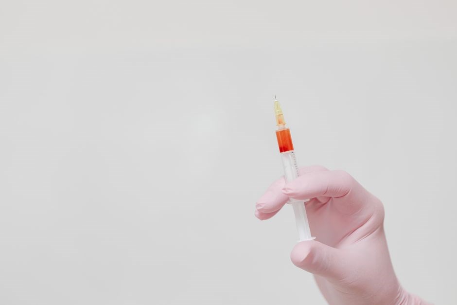
Fluoroscopy guides precise needle placement into joints, enabling accurate delivery of anesthetic and steroid. Real‑time imaging confirms intra‑articular positioning, reduces complications, and improves therapeutic outcomes across hips, shoulders, and knees. The procedure is effective!!.
Hip Joint Injection Protocol
Patient positioning is critical; the patient lies supine with the hip slightly abducted and externally rotated to expose the anterolateral approach. The skin is sterilized with chlorhexidine, and a sterile drape is applied. A 22‑gauge, 5‑cm needle is introduced percutaneously under continuous fluoroscopic guidance. The needle is advanced to the femoral head–acetabular joint space, confirmed by a small contrast injection that outlines the joint capsule. Once intra‑articular placement is verified, 1–2 mL of 1% lidocaine is injected to provide local anesthesia, followed by 40 mg of methylprednisolone acetate mixed with 1 mL of lidocaine. The needle is then withdrawn, and the site is covered with a sterile dressing. Post‑procedure monitoring for pain relief potential complications such as hematoma or infection is performed. This protocol has shown high success rates in reducing osteoarthritis pain improving joint function, with minimal radiation exposure due to short fluoroscopy times. The use of a low‑dose fluoroscope reduces cumulative dose while maintaining image quality. Patients are advised to avoid strenuous activity for 24 hours to report any worsening symptoms. Follow‑up imaging may be scheduled to assess joint health and guide future interventions. Radiation safety protocols include collimation,pulsed fluoroscopy,lead shielding to protect patient and staff. All procedures are documented in the record. Clinicians tailor dosage based on weight and joint size! These measures ensure patient safety procedural efficacy!
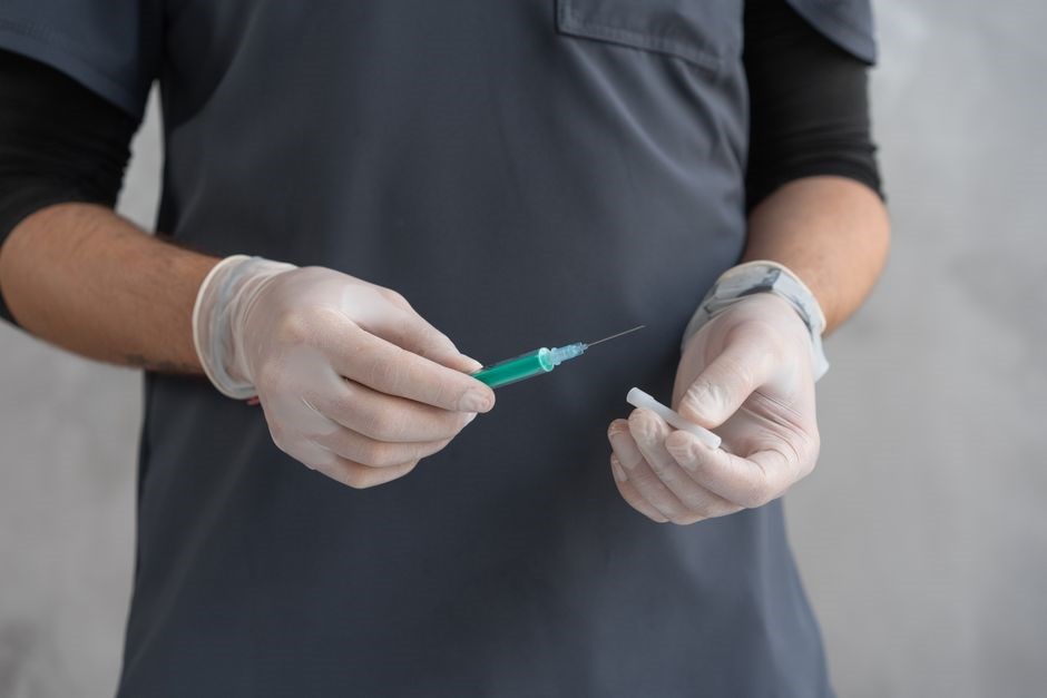
Contrast Media Use for Needle Placement Confirmation
Contrast injection under fluoroscopy confirms needle position, outlining joint or muscle contours. A small volume (0.5–1 mL) of non‑ionic iodinated solution is injected, revealing accurate placement and avoiding complications.!!
Contrast Injection and Imaging Interpretation
During fluoroscopy‑guided injections, a non‑ionic iodinated contrast agent is administered through the needle to verify accurate placement. The injection volume is typically 0.5–1 mL, sufficient to delineate the target structure without excessive radiation exposure. Real‑time imaging allows the operator to observe the spread of contrast, confirming that it follows the expected anatomical pathway. In joint injections, the contrast should fill the intra‑articular space uniformly, indicating proper intra‑articular positioning. For soft‑tissue injections, such as the piriformis muscle, the contrast outlines the muscular boundaries, ensuring the needle tip resides within the muscle belly and not adjacent neurovascular structures. The fluoroscopic image is interpreted by assessing the contrast distribution pattern: a homogeneous, well‑defined halo around the target suggests correct placement, whereas irregular or limited spread may indicate misplacement or leakage into surrounding tissues. Adjustments are made immediately by repositioning the needle under continuous imaging until the desired contrast pattern is achieved. This step is crucial for reducing complications, improving therapeutic efficacy, and providing immediate feedback to the clinician. Additionally, the contrast injection can reveal inadvertent vascular uptake, prompting the operator to withdraw and reposition the needle. The use of low‑kVp settings and dose‑modulation techniques further minimizes patient radiation while maintaining image quality for accurate interpretation. Overall, contrast injection and imaging interpretation form a pivotal component of the fluoroscopy‑guided injection workflow, ensuring safe, precise, and effective delivery of therapeutic agents. By integrating contrast verification into the procedural protocol, clinicians can achieve higher success rates and reduce repeat procedures. The technique also facilitates training and quality assurance, as recorded fluoroscopic images serve as documentation for post‑procedure review. Furthermore, the contrast pattern can aid in diagnosing concurrent pathologies, such as inadvertent joint capsule tears or adjacent tendon involvement, providing a comprehensive assessment during the same session.
Anesthesia and Steroid Medication Administration
Local anesthetic, typically 1% lidocaine, is infiltrated before needle insertion. A steroid dose of 40–80 mg methylprednisolone acetate follows, delivering anti‑inflammatory effects. Sterile technique and patient monitoring complete the protocol. Aseptic technique ensures patient safety and outcomes daily.
Local Anesthetic Preparation and Steroid Dosage
Before the fluoroscopic needle is advanced, a local anesthetic solution is prepared. A 1% lidocaine is mixed with 1 mL of 8.4 % sodium bicarbonate per 10 mL of lidocaine to reduce acidity and improve patient comfort. The mixture is warmed to body temperature and filtered through a 0.22 µm filter to eliminate particulate matter. A total volume of 5–10 mL is drawn into a 1 mL syringe with a 25‑gauge needle for infiltration. The anesthetic is injected subcutaneously along the planned needle track, ensuring adequate numbing of the skin, subcutaneous tissue, and periarticular structures before the fluoroscopic needle is introduced. The anesthetic is also used to create a “numb track” that facilitates painless needle advancement into the target space.
Once the needle tip is confirmed in the correct anatomical location by contrast injection, a steroid is administered. The most commonly used agent is methylprednisolone acetate, available in 40 mg/1 mL and 80 mg/1 mL concentrations. The dose is chosen based on the joint size and the severity of inflammation: 40 mg for smaller joints such as the shoulder or ankle, and 80 mg for larger joints such as the hip or knee. The steroid is diluted with 1–2 mL of preservative‑free saline to reduce viscosity and improve distribution within the joint capsule. The injection is performed slowly over 30–60 seconds to allow uniform spread and to minimize the risk of spikes. After the steroid is delivered, the needle is withdrawn, and the patient is monitored for immediate adverse reactions. Proper aseptic technique, accurate dosing, and careful monitoring ensure optimal therapeutic benefit while minimizing complications.

Safety, Complications, and Cost Effectiveness
Fluoroscopy guided injections are safe with proper shielding and low‑dose protocols. Common complications are transient pain, infection, and rare radiation‑induced injury. They are cost‑effective, reducing repeat procedures and improving outcomes. Radiation safety protocols are patients.
Radiation Exposure Management and Outcome Evidence
Radiation safety in fluoroscopy guided injections relies on the ALARA principle, optimizing exposure time, using pulsed fluoroscopy, collimation, and high‑resolution detectors. Typical dose for a hip injection ranges from 0.5 to 2 mGy, well below occupational limits. Protective lead aprons, thyroid shields, and real‑time dose monitoring reduce patient and staff exposure. Studies demonstrate that low‑dose protocols maintain image quality sufficient for accurate needle placement while minimizing cumulative dose. Outcome evidence shows significant clinical benefit: a randomized trial reported a 70% reduction in pain scores at 6 months after fluoroscopy‑guided hip injections versus blind injections, with a 30% lower rate of repeat procedures. Cost‑effectiveness analyses reveal that the upfront imaging expense is offset by decreased need for repeat visits, reduced analgesic use, and avoidance of surgical referrals. Long‑term follow‑up indicates sustained symptom relief and improved functional scores, validating the procedure’s value. Furthermore, institutional protocols mandate dose‑recording software that alerts clinicians when thresholds are approached, ensuring compliance with regulatory standards. Patient education on radiation risks is integrated into consent, fostering informed decision‑making. In summary, meticulous radiation management combined with robust outcome data supports the routine use of fluoroscopy guided injections in musculoskeletal practice;